| Issue |
BIO Web Conf.
Volume 24, 2020
International Conferences “Plant Diversity: Status, Trends, Conservation Concept” 2020
|
|
|---|---|---|
| Article Number | 00017 | |
| Number of page(s) | 5 | |
| DOI | https://doi.org/10.1051/bioconf/20202400017 | |
| Published online | 21 September 2020 | |
In Vitro Rhizogenesis of The Lavandula angustifolia Cultivars
Federal State Funded Institution of Science “The Labour Red Banner Order Nikita Botanical Gardens – National Scientific Center of the RAS”, Plant Developmental Biology, Biotechnology and Biosafety Department, 298648 Yalta, Russian Federation
* Corresponding author: This email address is being protected from spambots. You need JavaScript enabled to view it.
Abstract
In production, shoot cuttings of lavender are most often used for vegetative propagation, however, this method does not promote high propagation rate. The most effective method is propagation by axillary or terminal buds that requires further plant rooting in vitro. Due to, objective of our work was analysis of lavender in vitro rhizogenesis. Investigation was performed on Lavandula angustifolia cultivars with different concentration of the growth regulators in medium. After18 Day, Only 1 mg/l NAA did not stimulate rhizogenesis for ‘Record’ and ‘Sineva’ cultivars. Root primordia formed from the cambium cells. Further root growth accompanied by cleavage of the core tissues and they appeared on the shoot surface. Morphologically, a root cap, meristem, elongation zone and zone with root hairs were identified in de novo formed root in vitro. Along with normally roots, the appearance of roots accreted along their periphery with free apexes and roots with the decreased meristem was also noted. For ‘Prima’ cultivar, highest values of the mitotic index were observed on the hormone-free half-strength MS medium and on ½MS medium with 1.0 mg/l NAA. Thus, our data showed that root morphogenesis for lavender cultivars depended on the plant material and culture medium.
© The Authors, published by EDP Sciences, 2020
 This is an Open Access article distributed under the terms of the Creative Commons Attribution License 4.0, which permits unrestricted use, distribution, and reproduction in any medium, provided the original work is properly cited.
1 Introduction
In vitro tissue culture method allows to obtain plants from individual cells, tissues and organs on culture media. The basis of this method is the unique characteristic of plant cells – totipotency, that is mean the cell ability to realize their genetic information and give rise to a new organism. Under certain conditions, plant cells have differentiated in a second time and under the influence of external factors one or another way of morphogenesis has chosen. Morphogenesis is a complex process and its regulation is carried out at the cellular, tissue and organismal levels by factors such as light intensity, temperature, gas environment [1], nutrient composition, type of explant, its orientation on the medium [2, 3] and internal hormonal balance of explant [4]. These all determine the processes of division, elongation, differentiation, aging and cell death [5]. Essential oil plants are a renewable source of raw materials used in medicine and for the production of perfumes and cosmetics [6]. In the Crimea region, among the essential oil plants of Lamiaceae family, lavender is one of the most cultivated [7]. In production, vegetative propagation of lavender is most often used by shoot cuttings, however, this method does not promote high propagation rate [8, 9]. According to literature, the most effective method is propagation by axillary or terminal buds [10-12]. So there is necessity for fast and more effective rooting method in vitro for lavender plants. Thus, objective of this investigation was to study rhizogenesis for lavender on different growth culture media.
2 Materials and Methods
Lavender plants (Lavandula angustifolia Mill.) of the ‘Prima’, ‘Record’ and ‘Sineva’ cultivar grown ex situ at the collection plot of the Nikita Botanical Gardens were used for the investigation. Apical part of the shoots and auxiliary buds were cut, sterilized in SC2 biosafety cabinet (ESCO, Singapore) with 70% ethanol for 1 min, 0.3-0.4% Dez Tab solution with Tween (2-3 drops), cefotaxime for 30 min and transferred on the modified MS culture medium containing 0.7 mg/l BAP (6-benzylaminopurine, Sigma, USA) + 0.15 mg/l NAA (α-naphthylacetic acid, Sigma, USA) and cultured in phytochambers at 25±1°C under 16-h photoperiod and 37.5 μM-m-2-s-1 light intensity. For rhizogenesis, microshoots were placed on half-strength MS media: hormone-free; with 0.5 mg/l IBA (indole-3-butyricacid, Sigma, USA) and NAA; 0.5 mg/l IBA; 1 mg/l IBA and 1 mg/l NAA; 1 mg/l IBA; 1 mg/l NAA. Microshoots were grown in “BIOTRON” chambers at 25±1°C under 16-h photoperiod and 37.5 μM-m-2-s-1 light intensity.
For anatomical studies, the bottom parts of microshoots were cut and processed on freezing microtome (MZ-2, Ukraine) equipped with a lab cooling system OL-ZSO 30 (Inmedprom, Russian Federation). Sections were stained with methylene blue and investigated using a CX41 light microscope (Olympus, Japan) equipped with SC 50 camera (Olympus, Germany) and CellSens Imaging Software version 1.17.
In vitro lavender roots were cut off from plants and immediately immersed in Carnoy’s fixative (alcohol, chloroform, glacial acetic acid (6: 3: 1)) [13]. The fixation time was 3 hours. After, the roots were rinsed by 70% alcohol and preserved in the same alcohol solution. Further, roots were stained with 2% acetocarmine, by immersing the material in a dye and heating until its boiling. The roots were transferred in 45% acetic acid on the slide, meristem with root cap was cut and additionally chopped with a razor blade. The cuttings were covered by cover glass and pressed by match for obtaining cell monolayer. The samples were studied by phase contrast (Fig. 1) at magnification *400 using a CX41 microscope (Olympus, Japan) equipped with an SC 50 camera (Olympus, Germany) and CellSens Imaging Software version 1.17. Mitotic cells were counted on 5 digital images from each root in UTHSCSA ImageTool v. 3.00. Statistical analysis was performed using PAST software [14].
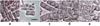 |
Fig. 1 Cell cycle phases of the lavender roots in vitro: a – interphase cells, b – prophase, c -metaphase, d – anaphase, e – telophase (acetocarmine, phase contrast). Bars – 20 μm. |
3 Results and discussions
Rhizogenesis in vitro is a complex process, including biochemical, histological and physiological changes. Root formation in vitro can occur on the plant vegetative organs as well as on their individual parts [15] and according to G.-J. De Klerk (1997) consists of several phases: acquisition of competence, induction and realization. The first phase corresponds to the cell differentiation; the second is sensitive to organogenic factors, such as growth regulators, which determine the formation of individual organs and are critical during this period; third is the deployment of a differentiation program [16]. In lavender plants spontaneously rooting was occurred on multiplication culture medium [17]. However, the ratio of auxins and cytokinins is important for rhizogenesis. At higher concentrations of cytokinins relative to auxins, the formation of shoots or buds occurs. Adding of the indole-3-acetic acid (IAA or its synthetic analogues – IBA and NAA in different concentration induces root formation [15]. Two last growth regulators were used in our study.
Visible root formation was observed on third day for ‘Prima’ cultivar. All media promoted root morphogenesis. On six day, microshoots of ‘Record’ cultivar had roots on four media, except 0.5 mg/l IBA and 1 mg/l NAA. ‘Sineva’ cultivar formed roots on medium with 0.5 mg/l IBA. After 18 Day, only 1 mg/l NAA was not stimulated rhizogenesis for ‘Record’ and ‘Sineva’ cultivars. Anatomically (Fig. 2), root primordia were formed from the morphogenic loci after cambium cell divisions in the internodes and nodes without a certain regularity, that was carried out likely due to the presence of competent cells. Their growth was accompanied by cleavage of the core tissues and roots appeared on the shoot surface.
Morphologically, a root cap, meristem, elongation zone and root hair zone were identified in the formed root. Along with normally developed roots, the appearance of roots accreted along their periphery with free apexes and roots with the decreased meristems was also noted (Fig. 3).
A new cells most sensitive to environmental factors were produced by meristem. The new cells form due to parent cell divisions, i.e. mitotic activity. The duration of the mitotic cycles of plants depends on the species characteristics of the plant, its age, temperature, aeration, moisture, light intensity, etc., and may also vary in plants of the same species [18].
Due to ‘Prima’ cultivar was characterized by fast root forming, compared to other cultivars, we investigated mitotic index (MI) of de novo formed root in vitro.
Highest values of the MI were observed on the hormone-free ½MC medium, as well as on ½MS medium with 1.0 NAA (Table). Other media showed a tendency to a slight decrease of MI. Mitotic activity is determined by the number of cells entering to mitosis per time, has rhythmic in which an increase of mitosis value is turned by a lower value and vice versa [19-21]. The apical meristems of various organs have their own number of mitoses varies depending on different factors [19] among which culture media with different concentration of growth regulators. For example, in vitro roots of Ocimum basilicum L. (var. Greek basil) on media supplemented with IBA (2 ml/l) and BAP (1 ml/l)+IAA (0.5 ml/l) had significantly increased MI. Media with NAA (2 ml/l), BAP (2 ml/l), BAP (0,2 ml/l) reduced MI, compared with control (complete MS medium without growth regulators) [22].
 |
Fig. 2 In vitro morphogenesis of the lavender roots: a – root formation in the node, b – root formation in the internode. xy – xylem, ph – phloem, c – cambium, lt – leaf trace, rt – root (light microscopy, methylene blue). |
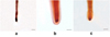 |
Fig. 3 Apexes of the roots, de novo formed in vitro on the lavender microshoots: a – normal root, b -roots accreted along their periphery with free apexes, c – roots with the decreased meristem was also noted. Scale Bar – 1 mm (light microscopy, acetocarmine). |
Mitotic index values of the lavender roots in vitro on media with different content of growth regulators.
4 Conclusions
Investigation of in vitro rhizogenesis of the lavender cultivars showed that plant material i.e. different genotype as well as growth regulator type are necessary factors for root formation. Competent cells promoting root appearance are cambial tissue. De novo formed roots in vitro had normal and deviated morphology. It was observed that mitotic index also depends on culture medium.
This study was funded by a research grant № 19-76-00023 of the Russian Science Foundation and done on the base of the Unique Scientific Installation “PHYTOBIOGEN” of the FSFIS “NBG-NSC”.
References
- K.W. Hughes, Env. Exp. Bot., 21, 281 (1981) [CrossRef] [Google Scholar]
- P. Bhatia, N. Ashwath, D.J. Midmore, In vitro Cell. Dev. Biol. Plant, 41, 457 (2005) [Google Scholar]
- P. Mazumdar, South Afr. J. Bot., 76, 337 (2010) [CrossRef] [Google Scholar]
- A. Garcia-Luis, Y. Bordon, J.M. Moreira-Dias, R.V. Molina, J.L. Guardiola, Ann. Bot., 84, 715 (1999) [Google Scholar]
- A.V. Babikova, T.Yu. Horpenchenko, Yu.N. Zhuravlev, Komarov readings, 55, 184 (2007) [Google Scholar]
- Z.G. Abbasova, Z.A. Mamedova, R.M. Mamedov, Plant chemistry, 1, 121 (2009) [Google Scholar]
- A.A. Pegushina, KANT, 2, 97 (2017) [Google Scholar]
- O.B. Skipor, A.V. Mishnev, Scientific Bulletins of BelSU, 33, 52 (2015) [Google Scholar]
- N.A. Yegorova, I.V. Mitrofanova, V.A. Brailko, О.А. Grebennikova, А.Е. Palii, I.V. Stavtseva, Russ. J. Plant Physiol., 66, 137 (2019) [Google Scholar]
- S. Gonçalves, A. Romano, Biotechnol. Adv., 31, 166 (2013) [CrossRef] [PubMed] [Google Scholar]
- D. Nicuţă, SCSB, 26, 19 (2017) [Google Scholar]
- I.V. Mitrofanova, A.E. Palii, O.A. Grebennikova, V.A. Brailko, N.P. Lesnikova-Sedoshenko, V.D. Rabotyagov, O.V. Mitrofanova, Agricultural Biology, 53, 539 (2018) [Google Scholar]
- Z.P. Pausheva, Practical work on Plant Cytology (Kolos, Moscow, 1980) [Google Scholar]
- Ø. Hammer, D.A.T. Harper, P.D. Ryan, Palaeontol. Electron., 4, 1 (2001) [Google Scholar]
- K.Yu. Guseva, I.D. Borodulina, E.P. Myakisheva, O.K. Tavartkiladze, News of AltSU 3, 56 (2013) [Google Scholar]
- G.J. De Klerk, B. Arnholdt-Schmitt, R. Lieberei, K-H. Neumann, Biol. Plant., 39, 53 (1997) [CrossRef] [Google Scholar]
- I.V. Mitrofanova, S.N. Chirkov, N.P. Lesnikova-Sedoshenko, S.V. Chelombit, A.V. Zakubanskiy, V.D. Rabotyagov, O.V. Mitrofanova, Acta Hortic., 1187, 37 (2017) [Google Scholar]
- S.A. Dmitrieva, F.V. Minibayeva, L.K. Gordon, Tsitologiia, 48, 475 (2006) [PubMed] [Google Scholar]
- A. Kunakh, Biopolym. Cell, 15, 343 (1999) [CrossRef] [Google Scholar]
- O.A. Artemenko, Space Sci. Technol., 23, 66 (2017) [Google Scholar]
- S.A. Bezhdugova, E.M. Gidova, M.M. Bittueva, T.Kh. Khandokhov, Z.I. Bogotova, M.N. Sitnikov, A.Yu. Paritov, G.Kh. Kirzhinov, T.Kh. Goguzokov, IJFAR, 3, 96 (2018) [Google Scholar]
- D-E. Maftei, Studia UBB Biologia, 59, 71 (2014) [Google Scholar]
All Tables
Mitotic index values of the lavender roots in vitro on media with different content of growth regulators.
All Figures
|
Fig. 1 Cell cycle phases of the lavender roots in vitro: a – interphase cells, b – prophase, c -metaphase, d – anaphase, e – telophase (acetocarmine, phase contrast). Bars – 20 μm. |
| In the text | |
|
Fig. 2 In vitro morphogenesis of the lavender roots: a – root formation in the node, b – root formation in the internode. xy – xylem, ph – phloem, c – cambium, lt – leaf trace, rt – root (light microscopy, methylene blue). |
| In the text | |
|
Fig. 3 Apexes of the roots, de novo formed in vitro on the lavender microshoots: a – normal root, b -roots accreted along their periphery with free apexes, c – roots with the decreased meristem was also noted. Scale Bar – 1 mm (light microscopy, acetocarmine). |
| In the text | |
Current usage metrics show cumulative count of Article Views (full-text article views including HTML views, PDF and ePub downloads, according to the available data) and Abstracts Views on Vision4Press platform.
Data correspond to usage on the plateform after 2015. The current usage metrics is available 48-96 hours after online publication and is updated daily on week days.
Initial download of the metrics may take a while.