| Issue |
BIO Web Conf.
Volume 25, 2020
International Scientific Online-Conference “Bioengineering in the Organization of Processes Concerning Breeding and Reproduction of Perennial Crops” 2020
|
|
|---|---|---|
| Article Number | 02018 | |
| Number of page(s) | 6 | |
| Section | The Fundamental Basis of Breeding and Improving of Horticultural Crops and Grape Varieties | |
| DOI | https://doi.org/10.1051/bioconf/20202502018 | |
| Published online | 01 October 2020 | |
Cortical photosynthesis as a physiological marker for grape breeding: methods and approaches
Institute of Basic Biological Problems, FRC PSCBR RAS, Pushchino, Moscow Region, 142290, Russia
* Corresponding author: This email address is being protected from spambots. You need JavaScript enabled to view it.
Abstract
Photosynthesis occurring in chlorenchymal tissues of lignified branches of perennial plants (cortical photosynthesis) has a significant impact on their productivity and resistance to adverse environmental conditions, such as water deficiency and low temperatures. Cortical photosynthesis occurring under the outer bark of a lignified grape vine can become a convenient marker for breeding freeze-tolerant varieties. The following approaches can be undertaken to assess the functional state of the cortical photosynthetic apparatus: (1) analysis of the variable chlorophyll fluorescence parameters and (2) biochemical analysis of photosynthetic membrane preparations. To evaluate these approaches, in this work we have carried out the comparative analysis of characteristics of the cortical photosynthetic apparatus in grape varieties differing in freeze tolerance.
This work was supported by grant №18-04-00079 from the Russian Foundation for Basic Research.
© The Authors, published by EDP Sciences, 2020
 This is an Open Access article distributed under the terms of the Creative Commons Attribution License 4.0, which permits unrestricted use, distribution, and reproduction in any medium, provided the original work is properly cited.
1. Introduction
The ability to absorb light energy and convert it into the energy of chemical bonds is present not only in young green shoots, but also in lignified branches and trunks in tissues below the outer bark [1-5]. It has been established that photosynthesis in the chlorenchymal tissues of the inner cortex of lignified plant organs, so-called “corticular photosynthesis” (CP), contributes to the carbon balance of the plant, radial growth of the stem, accumulation of bud biomass and the formation of young leaves [6-7]. The work of the corticular photosynthetic apparatus (CPA) partially compensate for the decrease in the assimilatory activity of leaves, especially during the leafless period, herewith the contribution of CP to the total carbon balance can be significant [1]. Thus, the activity of CP can be an important component of the productivity of perennial plants.
Obviously, the role of CP increases under unfavorable environmental conditions. The oxygen released in the CP process prevents hypoxia and maintains respiration, which can be especially important for the survival of plants at low temperatures during long winters. The cells of the wintering vine contain a significant amount of reserve carbohydrates, and the presence of sufficient oxygen is a prerequisite for the utilization of these reserve nutrients. Freeze tolerance, like other types of resistance to environmental stress factors, is a quantitative trait, largely shaped by energetic and metabolic constituents. The absorption of light energy, electron transport within photosynthetic electron-transport chain, and the re-fixation of carbon dioxide released during respiration in the inner cortex of perennial plants can be described using quantitative characteristics. Thus, the structural and functional characteristics of the cortical photosynthetic apparatus can become a convenient marker for the selection for highly productive and freeze tolerant grape varieties. Currently, the use of such markers is hampered due to a lack of sufficient knowledge about the structural organization and functional characteristics of CPA. The lack of reliable methods and developed protocols for CPA characterization also delays the implementation of this approach in practice. In this work, we have analyzed the parameters of variable chlorophyll fluorescence of intact vine tissues to compare the functional characteristics of the cortical photosynthetic apparatus in vine plants contrasting in freeze tolerance. In addition, we have developed a protocol for isolation of photosynthetic membranes from vine chlorenchyma suitable for biochemical studies.
Molecular mechanisms of freeze tolerance of grape plants remain poorly understood, and the present work is aimed at developing a new approach to improve freeze tolerance of this important agricultural crop [8, 9].
2. Materials and methods
The objects of the study were green and lignified same-year shoots of two hybrid grape varieties: interspecific hybrid of V. vinifera and V. amurensis - TANA42 (Kurchanskiy), Muskat kubanskiy (Precose Blanc × Muscat Hamburg) × Saperavi severnyy (Severnyy (Maleurengr seedling) × V. amurensis) × Saperavi), and an intraspecific hybrid of V. vinifera - TANA 33, Antaris (Saperavi × Tsimlyanskiy chernyy) × Krasnostop anapskiy (Krasnostop zolotovsky clone), grafted onto the Berlandieri × Riparia Kober 5BB rootstock. The plants were grown on the experimental plot of the North Caucasian Regional Research Institute of Horticulture and Viticulture.
Measurements of the parameters of variable chlorophyll fluorescence under laboratory conditions were carried out according to the method described earlier [10, 11]. The intensity of the acting light was 120 μmol photons s-1 m-2, λ≥625.
The procedure for isolation of chloroplasts and thylakoid membranes from chlorenchymal tissues is schematically represented in Fig. 1. The solution for isolation of chloroplasts contained 50 mM HEPES, pH 7.5, 0.4 M sucrose, 15 mM NaCl, and a mixture of protease inhibitors (cOmplete, EDTA-free Protease Inhibitor Tablets, Roche) (buffer A). The medium for resuspension of chloroplasts and thylakoid membranes contained 50 mM HEPES, pH 6.9, 0.4 M sucrose, 15 mM NaCl, and a mixture of protease inhibitors (cOmplete, EDTA-free Protease Inhibitor Tablets, Roche) (buffer B). The thylakoid membrane fraction was purified by centrifugation in a sucrose gradient at 100,000 g. A layer of thylakoid membranes was located between the 1M and 2M sucrose layers.
The oxygen-evolving activity of the samples was measured according to the previously described method [12].
3. Results and discussion
To compare the characteristics of CPA in the vine of the TANA33 and TANA42 plants, we analyzed the parameters of variable chlorophyll fluorescence measured on intact tissues of the vine with the removed outer bark, as described in the Materials and Methods section. This method revealed significant differences between studied grape varieties [13, 14].
Plants of freeze tolerant cultivar TANA42 are characterized by a higher photochemical activity in the chlorenchymal tissues of the vine, which is confirmed by higher values of maximum quantum yield of photosystem 2 (PS2), (Fv/Fm), reflecting the maximum efficiency of PS2, and the effective quantum yield of PS2 (YII), reflecting the proportion of absorbed excitation energy used for photochemical reactions (Table 1). After the treatment of samples with low positive and negative temperatures, the difference in the parameters characterizing photosynthetic apparatus in the TANA42 and TANA33 plants still remains or even becomes more noticeable.
Also, in the work, the isolation of photosynthetic membranes from leaf tissues, green and lignified vines was carried out in order to develop a protocol that allows preparation of photosynthetic membranes that retain functional activity (the scheme of the procedure is shown in Fig. 1).
Isolation of photosynthetically active membranes from lignified tissue is a difficult task [15], but it is the most straightforward way to gain the opportunity to perform biochemical studies aimed at elucidating the organization of CPA and identifying differences in the structure of CPA in plants that are tolerant and sensitive to low temperatures. Experiments have shown that the method of tissue grinding (using a grinder, mortar with pestel or homogenizer) affects the functional activity of the isolated preparations, and the best results were obtained by grinding the tissues in a buffer using a mortar and pestle. Also, some difficulties were associated with the presence of a large amount of mucus in the samples, which is most likely due to the high starch content in the tissues. The presence of mucus interfered with the sedimentation of the preparations during centrifugation. Preliminary soaking, centrifugation after a large dilution of the samples, the use of osmotic shock and the use of enzymes (cellulase, pectinase) did not give the desired result. Rapid grinding and immediate centrifugation of the samples proved to be the most effective method. In general, it was much easier to isolate preparations from the freeze tolerant variety TANA42 than from TANA33.
The functional activity of the obtained fractions was assessed by measuring the variable chlorophyll fluorescence (parameter Fv/Fm) and oxygen-evolving activity. The results of the analysis of the measurements of the variable fluorescence of chlorophyll are presented in Table 1. The results show that not only the tissues of the outer cortex (cortex) contain functional chloroplasts, but also the tissues of the xylem. Unfortunately, the isolated preparations had low photochemical activity. Surprisingly, the Fv/Fm values measured on isolated chloroplasts were lower than those of purified thylakoid membrane preparations. It is possible that the isolated chloroplasts contain substances that inhibit photochemical reactions, and the destruction of chloroplasts with subsequent purification of the thylakoid membranes by centrifugation in a sucrose gradient allows to get rid of these substances. It should be noted that the studied objects contain significant amounts of lignin, tannins, the presence of which in the samples complicates the isolation of thylakoid membranes and pigment-protein complexes (Fig. 2).
Unfortunately, chloroplasts and preparations of thylakoid membranes isolated from lignified vines did not exhibit oxygen-evolving activity, although preparations isolated from leaves and green vines actively evolve oxygen in the light. The rate of oxygen evolution by preparations of thylakoid membranes isolated from leaves was 124 μmol О2 per mg chlorophyll per hour, from green vine - 107 μmol О2 per mg chlorophyll per hour, i.e. the activity of preparations from green vine is comparable to preparations from leaves. Analysis of preparations isolated from leaves and lignified vines was carried out using electrophoresis in native polyacrylamide gel (PAGE) [16]. The analysis showed that the pigment-protein complexes of the analyzed samples differ markedly. The vine sample shows a band characteristic of the light-harvesting complex, and several bands characteristic of high-molecular-weight complexes, but the molecular weights of these complexes differ from those found in the samples obtained from the leaves.
Thus, the analysis of the variable chlorophyll fluorescence of intact vine tissues is a convenient method for assessing the functional activity of CPA, which can be used to identify plants with high CP activity at an early stage in breeding practice. The methods of isolation of functional preparations of photosynthetic membranes from lignified vines and analysis of pigment-protein complexes of CPA require further optimization. The successful solution of these problems will help to identify biochemical markers associated with the activity of CPA, applicable in breeding practice.
Comparative analysis of the characteristics of CPA in vine tissues and isolated photosynthetic membranes.
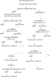 |
Fig. 1. Isolation of photosynthetic membranes from vine chlorenchyma * - collecting samples for further analysis. |
 |
Fig. 2. Electropherogram of thylakoid membranes isolated from the leaf and vine. The arrows indicate the bands corresponding to the light-harvesting complexes of PS2. |
References
- A. Saveyn, K. Steppe, N. Ubierna, T.E. Dawson, Plant Cell Environ, 33(11), 1949–1958 (2010) https://doi.org/10.1111/j.1365-3040.2010.02197.x [CrossRef] [PubMed] [Google Scholar]
- K.G. Tikhonov, M.S. Khristin, V.V. Klimov, M.A. Sundireva, V.D. Kreslavski, R.A. Sidorov, V.D. Tsidendambayev, T.V. Savchenko, Russ. J. Plant Physiol., 64(1), 73–82 (2017) https://doi.org/10.1134/S102144371606011X [Google Scholar]
- J. Liu, L. Gu, Y. Yu, G. Ju, Z. Sun, Forests, 9(4), 207 (2018) https://doi.org/10.1134/S102144371606011X [Google Scholar]
- D. Johnstone, M. Tausz, G. Moore, M. Nicolas. AoB Plants., 6, plt057 (2014). https://doi.org/10.1093/aobpla/plt057 [Google Scholar]
- S. Asao, R. Bedoya-Arrieta, M.G. Ryan, Tree Physiol., 35(2), 148–159 (2015) http://doi.org/10.1093/treephys/tpu107 [CrossRef] [PubMed] [Google Scholar]
- A. Saveyn, K. Steppe, N. Ubierna, T.E. Dawson, Plant Cell Environ, 33(11), 1949–1958. (2010) https://doi.org/10.1111/j.1365-3040.2010.02197.x [CrossRef] [PubMed] [Google Scholar]
- Y.Y. Hu, Y.L. Zhang, H.H. Luo, W. Li, R. Oguchi, D.Y. Fan, W.S. Chow, W.F. Zhang, Planta, 235(2), 325–336 (2012) https://doi.org/10.1007/s00425-011-1511z [CrossRef] [PubMed] [Google Scholar]
- T.N.L. Grant, I.E. Dami, Am. J. Enol. Viticult., 66(2), 195–203 (2015) http://doi.org/10.5344/ajev.2014.14101 [CrossRef] [Google Scholar]
- G.R. Strimbeck, P.G. Schaberg, C.G. Fossdal, W.P. Schroder, T.D. Kjellsen Front. Plant Sci., 6, 884 (2015) http://doi.org/10.3389/fpls.2015.00884 [CrossRef] [PubMed] [Google Scholar]
- D. Yanykin, M. Sundyreva, A. Khorobrykh, G. Semenova, T. Savchenko Biochim. Biophys. Acta. Bioenerg., 1861(11), 148–260 (2020) http://doi.org/10.1016/j.bbabio.2020.148260 [Google Scholar]
- MULTI-COLOR-PAM Manual, 3rd Edition (Heinz Walz GmbH, 106–110, 2013) https://www.walz.com/downloads/manuals/multi-color-pam/MC_PAM06.pdf [Google Scholar]
- T. Savchenko, D. Yanykin, A. Khorobrykh, V. Terentyev, V. Klimov, K. Dehesh, Planta, 245(6), 1179–1192 (2017) https://doi.org/10.1007/s00425-017-2674-z [CrossRef] [PubMed] [Google Scholar]
- M. Brestic, M. Zivcak, In: A.B. Das, G.R. Rout (eds), Molecular stress physiology of plants (Springer, New Dehli, 87–131, 2013) https://doi.org/10.1007/978-81-322-0807-5_4 [CrossRef] [Google Scholar]
- H.M. Kalaji, G. Schansker, M. Brestic, F. Bussotti, A. Calatayud, L. Ferroni, V. Goltsev, L. Guidi, A. Jajoo, P. Li, P. Losciale, V.K. Mishra, A.N. Misra, S.G. Nebauer, S. Pancaldi, C. Penella, M. Pollastrini, K. Suresh, E. Tambussi, M. Yanniccari, M. Zivcak, M.D. Cetner, I.A. Samborska, A. Stirbet, K. Olsovska, K. Kunderlikova, H. Shelonzek, S. Rusinowski, W. Bąba, Photosynth. Res., 132, 13–66 (2017) https://doi.org/10.1007/s11120-016-0318-y [CrossRef] [PubMed] [Google Scholar]
- D. Holá, M. Kočová, O. Rothová, E. Hlízová, L. Fridrichová, Z. Lhotáková, J. Albrechtová, Photosynthetica 50(2), 291–304 (2012) https://doi.org/10.1007/s11099-012-0028-4 [Google Scholar]
- S. Järvi, M. Suorsa, V. Paakkarinen, E.M. Aro, Biochem J., 439(2), 207–214 (2011) http://doi.org/10.1042/BJ20102155 [CrossRef] [PubMed] [Google Scholar]
All Tables
Comparative analysis of the characteristics of CPA in vine tissues and isolated photosynthetic membranes.
All Figures
|
Fig. 1. Isolation of photosynthetic membranes from vine chlorenchyma * - collecting samples for further analysis. |
| In the text | |
|
Fig. 2. Electropherogram of thylakoid membranes isolated from the leaf and vine. The arrows indicate the bands corresponding to the light-harvesting complexes of PS2. |
| In the text | |
Current usage metrics show cumulative count of Article Views (full-text article views including HTML views, PDF and ePub downloads, according to the available data) and Abstracts Views on Vision4Press platform.
Data correspond to usage on the plateform after 2015. The current usage metrics is available 48-96 hours after online publication and is updated daily on week days.
Initial download of the metrics may take a while.