Figure 2.
Download original image
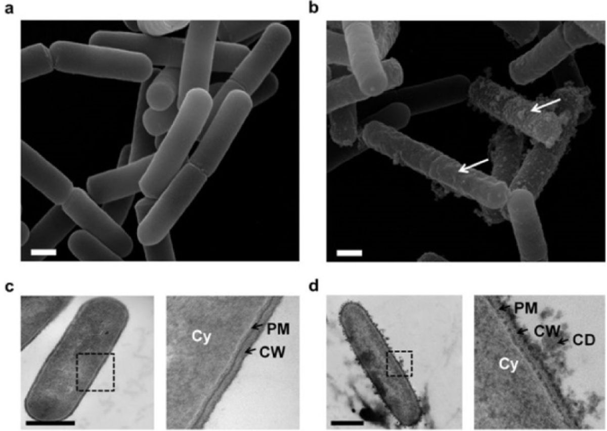
(a) SEM image showing population of untreated bacteria. Bacteria are smooth. (b) Pulsed bacteria were visualized by SEM after PEF exposure (1000 micropulses at 7.5 kV/cm). White arrows indicate surface damage. (c) TEM image of untreated Bacillus pumilus. Inset shows bacterial architecture with the cytoplasm (Cy), plasma membrane (PM) and cell wall (CW). (d) TEM image of a pulsed bacterium after PEF. Inset shows the expulsion of cell-debris (CD), damage to the PM and the CW. Scale bars: 500 nm. Flavien Pillet et al 2015 – Université de Toulouse.
Current usage metrics show cumulative count of Article Views (full-text article views including HTML views, PDF and ePub downloads, according to the available data) and Abstracts Views on Vision4Press platform.
Data correspond to usage on the plateform after 2015. The current usage metrics is available 48-96 hours after online publication and is updated daily on week days.
Initial download of the metrics may take a while.